
Why Midwives Don’t Perform Routine Pregnancy Scans & When We Refer
- Samantha Pieterse

- Jul 29, 2025
- 8 min read
Updated: Feb 11

From the moment you see those two lines on a pregnancy test, reassurance becomes everything. It’s completely natural to want to see your baby, hear the heartbeat, and know that everything is progressing well.
Ultrasound scans can provide that reassurance, but they’re not needed at every visit (if you don't want one). While it’s allowed to have a scan more frequently, more scans don’t automatically mean better care. What matters most is timing them at the stages where they provide meaningful medical information.
In South Africa, most midwives do not perform ultrasound scans in-clinic. Pregnancy scans are usually done by trained sonographers or radiology practices. As midwives, our role is to guide you on when scans are necessary, refer you appropriately, and then interpret the results within the bigger picture of your pregnancy.
Let’s walk through when scans are recommended and how you can go about them if you are receiving midwife care for your pregnancy.
Curious about what your first check-up looks like? Here’s what we do at your first pregnancy visit.
8 Weeks: Confirmation of Pregnancy & Ectopic Check
The first scan we usually recommend is around 8 weeks of pregnancy. At this stage, the most important question isn’t just, “Is there a heartbeat?” It’s: “Is the pregnancy in the right place?”
An ectopic pregnancy happens when a fertilised egg implants outside the uterus, most commonly in the fallopian tube. Because the fallopian tube cannot safely stretch to support a growing pregnancy, an ectopic pregnancy can become serious if not diagnosed early. Confirming that the pregnancy is inside the uterus is therefore one of the most important reasons for this early scan.
We’re often asked: “Can I have a scan at 5 or 6 weeks just to check everything is okay?”
Those early weeks can feel incredibly long. But before 7–8 weeks, it is often too early to see much on ultrasound, even in a completely healthy pregnancy. At 5–6 weeks, a scan may only show a small gestational sac, and sometimes a yolk sac. The heartbeat typically becomes visible from around 6–7 weeks onward, depending on dating accuracy and equipment quality. If a heartbeat isn’t seen yet, it doesn’t automatically mean something is wrong. It may simply be too early.
Scanning too soon can sometimes create unnecessary anxiety and repeat appointments. By around 8 weeks, a sonographer can usually obtain clear confirmation and accurate dating in a single appointment.
The 8-week scan allows us to:
Confirm the pregnancy is inside the uterus
Rule out an ectopic pregnancy
Confirm a heartbeat
Estimate your due date based on your baby’s size
This scan provides important clarity in early pregnancy and helps us plan the months ahead with confidence.
It is particularly important if you’ve experienced:
Pain or bleeding
A previous miscarriage
A previous ectopic pregnancy
Irregular cycles
Uncertainty about your dates
As midwives, we refer you to a trusted sonographer for this scan. Once it’s completed, we review the report together and guide your ongoing care from there.

📍We refer our patients to LifeScan Studio for this scan. Their team is kind, gentle, and experienced with early pregnancies.
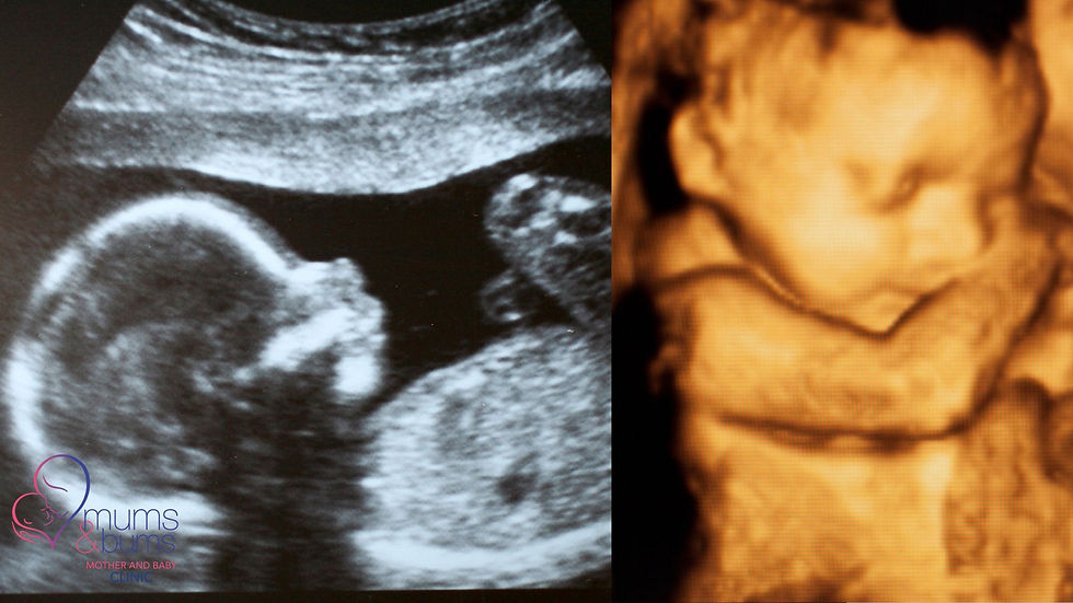
12 Weeks: Early Anomaly & Screening
Between 11 and 13 weeks, we recommend an early screening scan. This is often considered the first major developmental checkpoint in pregnancy. By this stage, your baby has grown enough for us to look beyond simply confirming a heartbeat. We’re now assessing how development is progressing.
This scan allows the sonographer to:
Measure your baby’s growth (crown-to-rump length)
Reconfirm or adjust your due date if needed
Assess early structural development (brain, spine, abdominal wall, limbs)
Measure the nuchal translucency
The nuchal translucency is a small, temporary fluid space at the back of your baby’s neck. Measuring this, together with your age and certain blood tests (if you choose), helps calculate the risk of chromosomal differences such as Down syndrome.
It’s important to understand that this is a screening test, not a diagnosis. It does not determine whether something is wrong. It simply helps identify pregnancies that may require further testing.
This scan is often combined with first-trimester blood screening or non-invasive prenatal testing (NIPT), depending on your preferences and risk factors.
The timing of this scan is important. The 11–13-week window is when measurements are most accurate for screening.
For many parents, this is also the scan where pregnancy begins to feel more tangible. Your baby is clearly formed, moving, and recognisably “baby-shaped.”
As with all imaging, we refer you to a qualified sonographer for this scan and then review the results together to ensure your ongoing care remains appropriate and reassuring.

This is a great time to start thinking about your birth prep. Check out our antenatal classes.
20 Weeks: Detailed Anatomy & Gender
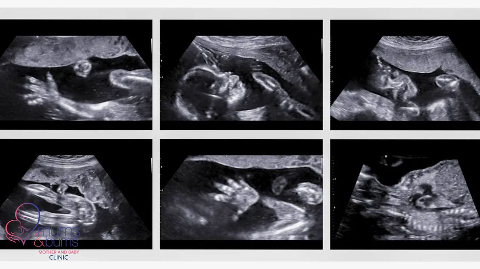
Around 20 weeks of pregnancy, we recommend what is often called the detailed anatomy scan. This is the most comprehensive ultrasound of your entire pregnancy. By this stage, your baby is large enough and developed enough for the sonographer to examine structures carefully, from head to toe.
During this scan, the sonographer will assess:
Brain structure and development
The four chambers of the heart and major vessels
Kidneys and bladder
Spine and limbs
Facial structures (including lips and nose)
Placenta location
Amniotic fluid levels
Umbilical cord insertion
Cervix length
The primary goal of this scan is to screen for major structural abnormalities and ensure that your baby’s growth is appropriate for gestational age.
It is important to remember that this is still a screening scan. Most results are reassuring. If anything appears unclear or requires further assessment, you may be referred for more specialised imaging.
Placenta location and cervix length are especially important at this stage. A low-lying placenta or a shortened cervix may require closer monitoring later in pregnancy, and identifying these early allows us to plan safely.
If you would like to know your baby’s gender, this is usually the stage when it can be identified, provided your baby cooperates.
After your scan, we review the report together and incorporate the findings into your pregnancy plan, ensuring that your care remains tailored and appropriate.
36–38 Weeks: Positioning
Not every pregnancy requires a late ultrasound.
By the third trimester, we can usually assess your baby’s position and growth through clinical examination, feeling your abdomen, measuring fundal height, and listening to the heartbeat. In many cases, this provides sufficient information without additional imaging.
However, we may recommend a scan between 36 and 38 weeks if clarification is needed.
This might include situations where:
Your baby’s position is unclear on examination
Your baby is measuring smaller or larger than expected
There are concerns about amniotic fluid levels
We need to confirm the placenta location
At this stage, the scan primarily helps confirm your baby’s position. Ideally, most babies are head-down (cephalic) by 36 weeks. Occasionally, a baby may be breech (bottom-down) or lying sideways (transverse). Knowing this allows us to discuss safe birth options and plan appropriately.
A late scan may also provide an estimated fetal weight, though it’s important to remember that these measurements are not exact.
If a scan is recommended, we refer you to a qualified sonographer and then review the findings together to ensure your birth plan remains safe and appropriate. In many pregnancies, however, no additional imaging is needed, and that is completely normal.
Why a Scan is Not Needed at Each Visit
Pregnancy care isn’t just about imaging. It’s about monitoring your health and your baby’s development. Routine pregnancy visits are designed to assess progress without over-medicalising what is, in most cases, a healthy and normal process.
At each appointment, we carefully monitor:
Your baby’s growth through abdominal measurement
Your baby’s heartbeat using a Doppler
Your blood pressure
Signs of preterm labour
Your symptoms and overall well-being
Your emotional health and preparation for birth
These clinical assessments provide meaningful information about how your pregnancy is progressing. Ultrasounds are valuable tools, but they are just that: tools. They complement care. They don’t replace it.
More scans don’t automatically provide more reassurance or better outcomes. What matters most is using ultrasound at the right time, for the right reason, and within the bigger picture of your pregnancy. In midwife-led care, we focus on balanced, evidence-based monitoring, ensuring you receive appropriate imaging when it’s needed, without unnecessary interventions.
Where Are Pregnancy Scans Done?
Mums & Bums does not perform ultrasounds in-clinic. We do, however, provide midwife-led pregnancy care in Centurion, guiding you on when scans are needed and referring appropriately.
In South Africa, pregnancy scans are typically done by qualified sonographers or radiology practices. When a scan is needed, we refer you to the appropriate facility and then review the results together as part of your ongoing pregnancy care.
This ensures you receive accurate imaging, alongside personalised midwife support and interpretation.
Recap: Pregnancy Milestones That May Require Imaging
Weeks | Purpose |
< 8 weeks | Usually, it's too early. Wait a bit. |
8–9 weeks | Confirm pregnancy, rule out ectopic, and determine the due date. |
12–13 weeks | Nuchal translucency, early anatomy. |
20 weeks | Full anatomy, gender reveal if possible. |
36–38 weeks | Check your baby’s position before birth (if needed). |
Let’s Keep You Reassured During Your Pregnancy
Pregnancy is full of questions, flutters, and decisions. Our job is to help you feel prepared and informed. At Mums & Bums Centurion, we:
Track baby’s growth and your health.
Help you time and book your ultrasounds.
Offer personalised pregnancy checkups, prenatal classes, and baby care under one roof.
👉🏻 Need guidance on your next scan? Book a pregnancy consultation here.
👉🏻 Want to prep for birth? Learn more about how our antenatal classes answer all your questions.
Questions About Your Pregnancy Timeline?
Whether you’re wondering when to book your scan or what’s really happening in week 13, we’re here to walk it with you.
💬 Friendly care, expert advice, and space to ask everything you’re thinking.
FAQ's
Do midwives perform pregnancy scans in South Africa?
Most midwives in South Africa do not perform ultrasound scans in-clinic. Pregnancy scans are usually done by qualified sonographers or radiology practices. Midwives refer for imaging when needed and then interpret the results as part of ongoing pregnancy care.
How many scans are medically necessary during pregnancy?
In an uncomplicated pregnancy, most women require around three key scans: an early confirmation scan (around 8 weeks), a screening scan at 12 weeks, and a detailed anatomy scan at 20 weeks. A late scan at 36–38 weeks may be recommended if clinically indicated.
Is it safe to have multiple pregnancy scans?
Ultrasound scans are considered safe when performed by trained professionals. However, more scans do not necessarily improve outcomes. What matters most is having scans at the right time for medical reasons.
Can I have a scan at every pregnancy visit?
You can have scans at every pregnancy visit, but it is not usually necessary in a healthy pregnancy. Routine midwife visits focus on clinical monitoring, and imaging is recommended only when it provides meaningful information.
When should I have my first pregnancy scan?
We typically recommend the first scan around 8 weeks. This confirms the pregnancy is in the uterus, checks for a heartbeat, and provides accurate dating.
What is an ectopic pregnancy?
An ectopic pregnancy occurs when a fertilised egg implants outside the uterus, most commonly in a fallopian tube. It cannot develop safely and requires medical attention. An early scan helps rule this out.
What if my baby is not head-down at 36 weeks?
If your baby is in breech or lying sideways near the end of pregnancy, we may refer you for a scan to confirm the position. This helps guide safe birth planning.


Comments